


The Nervous System
Teacher Pages
 The nervous system allows for sensing, response, and control. Response by the nervous system is accomplished by motor control. (Contrast this response with the chemical control used by the endocrine system.)
How does the nervous system accomplish this? How are messages transmitted throughout the organism and how are the messages interpreted? How does the organism respond?
Environmental (internal or external to the body) messages (stimuli) are received, transmitted, and interpreted by the functional unit of the nervous system—the neuron. Appropriate responses are carried out by effectors (a muscle or gland).
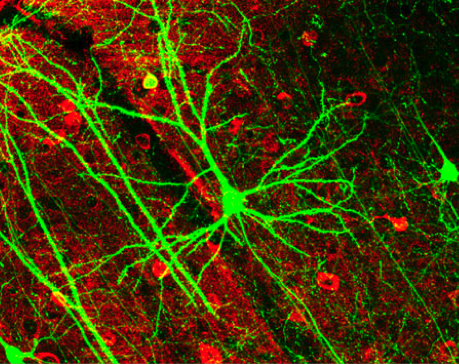
The Neuron
The neuron, a nerve cell, is the functional unit of the nervous system that carries the impulse (the message) to the appropriate part of the nervous system or interprets the impulse and allows a response. There are three types of neurons and each has different functions.
1) sensory neurons—receive impulses and carry them from the sense organs to the spinal cord or brain.
2) interneurons—connect sensory and motor neurons and interpret the impulse; only in the brain and spinal cord.
3) motor neurons—carry impulses from the brain and spinal cord to muscles or glands
 The Response Mechanism
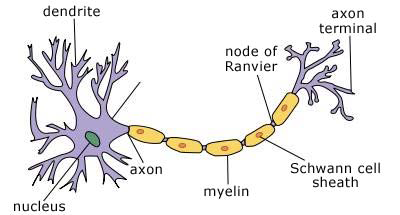
When a stimulus is received by a sensory neuron, the impulse (or message) is carried through fibrous extensions called dendrites to the cell body. The cell body is made up of cytoplasm, cytoplasmic structures, and a nucleus, which controls neuron function. The impulse travels through the cell body and is carried through the axon to the end brush, a collection of fibers that extend off the axon. Here, the impulse triggers a release of chemicals that allow the impulse to travel through the synapse—the space between the axon of one neuron and the dendrites of the next.
An impulse travels along the neuron pathways as electrical charges move across each neural cell membrane. Ions moving across the membrane cause the impulse to move along the nerve cells.
The difference in electrical charge on each side of the cell membrane (caused by differing numbers of positively and negatively charged ions) produces a resting potential. Neurons have a resting potential of approximately 70 millivolts (mV).
Specifically, the cell membrane proteins pump sodium ions (Na+) out of the neuron and pump potassium ions (K+) into the neuron. Active transport mechanisms and leaking back and forth of both the Na+ and K+ ions produce a negative charge on the inside of the neuron’s cell membrane.
 An impulse begins when a neuron is stimulated by another neuron or by a stimulus in the environment. The cell membranes begin to change the flow of ions and a reversal of charges, the action potential, results. An impulse that changes one neuron, changes the next. The impulse movement continues along the pathway in this way.
An impulse can travel quickly through the nervous system. Many vertebrates’ neurons, including humans, have several features that allow for maximum reactivity. Vertebrate axons have a myelin sheath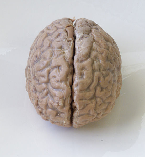 that allows for faster travel of the impulse. Nodes on the axons also allow impulses to jump from node to node instead of traveling through the entire axon. This also allows for faster response. The more quickly an organism can respond, the better adapted it is to its environment.
Nervous System Organization
Two main parts of the human nervous system are the:
1) Central nervous system—consists of the brain and spinal cord.
The brain and spinal cord are protected by three layers of tissue called the meninges. Between the layers is a space filled with cerebrospinal fluid. This fluid acts of a shock absorber to protect against injuries.
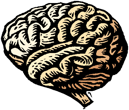
The Brain
Consisting of more than 100 billion neurons with a mass of approximately 1.4 kilograms, the brain is the organ by which we think, react, respond, and process what we know about our environment. Our sense organs receive stimuli from the environment, but can not process or interpret them.
Our brain has specialized centers for processing sensory information and for response. Therefore, in a real sense, we see with our brain, we hear with our brain, we taste with our brain, and so on. Our eyes may take in the stimuli, but without our brain, we would “see” nothing.
The brain is divided into regions that control different functions. Below is an abbreviated list of the regions and what they control. This list is not meant to be detailed enough for a comprehensive study of the brain, but it should help you to understand the way the brain is further specialized by area.
Cerebrum—site for intelligence, learning, and judgment, voluntary muscle control, storing information of memory, personality
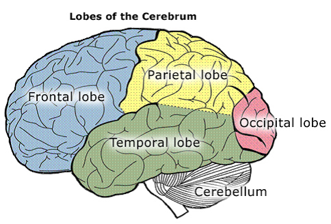
5 lobes and what they control:
1) frontal lobe—concentration, planning, problem solving
2) parietal lobe—understanding speech, using words
3) temporal lobe—interpretation of sensory stimuli, memory of visual and
auditory patterns
4) occipital lobe—visual interpretations, combining visual images with
recognition
Cerebellum—communicates with other parts of the central nervous system, interprets sensory information from the limbs, coordinates complex muscular movements, helps maintain posture
Brain stem—connects the cerebrum to the spinal cord.
1) midbrain—contains reflex centers, visual reflexes, auditory reflex centers
2) pons—transmits impulses to and from medulla oblongata, and cerebrum, relays messages from peripheral nerves to higher brain centers
3) medulla oblongata—control visceral activities
cardiac center—transmits impulses to the heart to control heart rate
vasomotor center—sends impulses to blood vessels to dilate or contract thereby altering blood pressure
respiratory center—regulates the rate, the rhythm, and depth of breathing
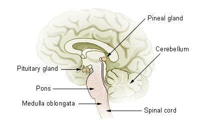
Spinal Cord—31 pairs of spinal nerves originate from the spinal cord to provide communication between the spinal cord and the limbs and trunk. The nerves are named for the level from which they come out. There are 8 pairs of cervical nerves, twelve pairs of thoracic nerves, five pairs of lumbar nerves, 5 pairs of sacral nerves, and one pair of coccygeal nerves.
2) Peripheral nervous system—consists of all the nerves except for the brain and spinal cord
Two main subdivisions:
- somatic nervous system—consists of the cranial and the spinal nerve fibers
- autonomic nervous system—neurons that connect the central nervous system to the viscera to control unconscious activities
Check for Understanding
- Explain the relationship between the central nervous system and the peripheral nervous system.
Both work together to sense internal and external stimuli. The central nervous system consists of the neurons in the brain and spinal cord. The peripheral nervous system consists of all other neurons throughout the body.)
- How are neurons classified according to their function?
Sensory neurons receive impulses and carry them from the sense organs to the spinal cord or brain. Interneurons connect sensory and motor neurons and interpret the impulse. Motor neurons carry impulses from the brain and spinal cord to muscles or glands.
- Draw and label the structure of a typical nerve cell.
- Explain how an impulse moves along a neural pathway.
An impulse travels along the neuron pathways as electrical charges move across each neural cell membrane. Ions moving across the membrane cause the impulse to move along the nerve cells.
The difference in electrical charge on each side of the cell membrane (caused by differing numbers of positively and negatively charged ions) produces a resting potential. Neurons have a resting potential of approximately 70 millivolts (mV).
Specifically, the cell membrane proteins pump sodium ions (Na+) out of the neuron and pump potassium ions (K+) into the neuron. Active transport mechanisms and leaking back and forth of both the Na+ and K+ ions produce a negative charge on the inside of the neuron’s cell membrane.
An impulse begins when a neuron is stimulated by another neuron or by a stimulus in the environment. The cell membranes begin to change the flow of ions and a reversal of charges, the action potential, results. An impulse that changes one neuron, changes the next. The impulse movement continues along the pathway in this way.
When the impulse reaches the end of one neuron (the axon), the impulse reaches a synapse. A synapse is the space between neurons. This space is filled with neurotransmitters, chemicals which allow the impulse to travel through the synapse to the next neuron.
5. Label the four lobes of the cerebrum and label the cerebellum on the following diagram.
6. Name the major portions of the brain and describe general functions of each. Complete the following table.
|
Brain section |
Function |
|
_Cerebrum_____ (has 4 lobes) |
|
|
1) parietal |
understanding speech, using words
|
|
2) occipital |
visual interpretations, combining visual images with recognition
|
|
3) temporal |
interpretation of sensory stimuli, memory of visual and auditory patterns
|
|
4) frontal |
concentration, planning, problem solving
|
|
_Cerebellum____ |
communicates with other parts of the central nervous system, interprets sensory information from the limbs, coordinates complex muscular movements, helps maintain posture
|
|
_Brain stem__(has 3 sections) |
connects the cerebrum to the spinal cord
|
|
1) _midbrain______
|
contains reflex centers, visual reflexes, auditory reflex centers
|
|
2) _pons__________
|
transmits impulses to and from medulla oblongata
|
|
3) _medulla oblongata_
|
control visceral activities
|
7. The strength of an impulse is constant; it is always the same. How, then, do we differentiate between strong or weak stimuli?
Strong stimuli activate more sensory neurons and are perceived as “stronger” even though each neuron is stimulated at the same level. Weak stimuli do not activate as many sensory neurons, and are therefore, perceived as weak.
8. What functional losses would you expect in a patient who has suffered injury to the right occipital lobe? To the right temporal lobe?
Functional losses after injury to the right occipital lobe include abnormalities in visual interpretations and combining visual images with recognition.
Functional looses after injury to the right temporal lobe include problems in interpretation of sensory stimuli and memory of visual and auditory patterns.
9. In neurological disorders such as multiple sclerosis, nerve fibers in the central nervous system lose their myelin sheaths. How would this affect control of skeletal muscle tissue?
Control of skeletal muscle tissue would be more difficult and take longer to accomplish.