


The Muscular System
Student Pages
The muscular system allows us to move. Adults have a certain fixed number of muscle cells, approximately 602. . Through exercise, such as weight lifting, the cells enlarge but the number of cells does not increase.
Structure and Function
There are three types of muscle tissue—smooth, skeletal, and cardiac. Each has its own distinctive function, and therefore, its own distinctive structure, but all muscles allow movement of some kind.
Skeletal Muscle
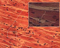
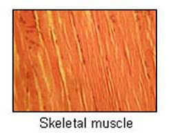 Skeletal muscle is called voluntary because you can control this type of muscle. You can voluntarily choose to move them. These muscles attach to bones, move the skeleton, and are found in the arms, legs, neck, or anywhere you can voluntarily move a body part. These muscles produce strong contractions.
Structurally, these muscle fibers appear striated (striped) when magnified, have more than one nucleus, and may be up to 30 cm long in humans. Look at the table in this lesson that compares the features of the three types of muscle tissues.
Smooth Muscle
 Smooth muscle is called involuntary muscle because you can not control this type of muscle movement. These muscles line internal organs, blood vessels, and organs such found in the digestive and reproductive systems.
Structurally, these muscle fibers appear non-striated (not striped) when magnified, have one nucleus per cell, and are usually short. They produce weaker contractions.
 Cardiac Muscle
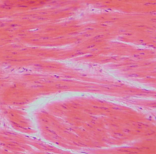
Cardiac muscle tissue is involuntary muscle and found, as its name would suggest, only in the heart. You can not control your heart muscle; it works automatically for you.
Structurally, these muscle fibers appear striated when magnified, have more than one nuclei per cell, and are also branched in appearance.
Muscle contraction mechanisms
How do muscles of any kind contract? What makes the muscle cells work together to move?
Muscle fibers are made up of alternating patterns of thick and thin filaments. The thick filaments are proteins called myosin; the thin filaments are proteins called actin. Other proteins are involved in the contraction process, but these two are the most important proteins for movement. In contraction, the end of one myosin molecule forms a “bridge” to an actin molecule. Using energy from cellular metabolic processes, the bridge changes shape and pulls at the thin actin molecule. The molecular bridge releases, then connects again to the thin filament to contract again. The cycle repeats and the muscle fibers shorten.
Hundreds of thousands of fibers shorten in this way and with force. This process is known as the sliding filament model of muscle contraction because the actin filaments slide over the myosin filaments.
The muscular system works with the nervous system to produce the contraction. Neurons (nerve cells) form connections to one or more muscle cells. A chemical, acetylcholine, is released when an impulse is sent which allows the myosin-actin bridge to form. The muscle cells remain in contraction until the release of acetylcholine stops and an enzyme destroys the remaining chemical.
If an impulse stimulates many neurons connected to muscle fibers, then the force of the contraction is large. If an impulse stimulates only a small number of neurons connected to a small number of muscle cells, then the contraction is weaker. In this way, we can control the force of motion that is necessary to move.
You should be more aware now of the interconnectedness of our body systems. How our body moves is an example of this dependency among systems. Our muscular system relies on our nervous system for the impulses necessary to produce movement. Our muscles must be connected to the skeletal system (bones) to allow for strength and movement and our circulatory system must transfer the chemicals necessary (by way of the digestive system) for the processes that provide biological energy.
The muscular system allows us to move. Adults have a certain fixed number of muscle cells, approximately 602. Through exercise, such as weight lifting, the cells enlarge but the number of cells does not increase.
A word about energy
As you recall from your studies of cellular respiration, energy for metabolic functions is supplied by “biological energy”, ATP. Cells produce ATP from glucose and other foods.
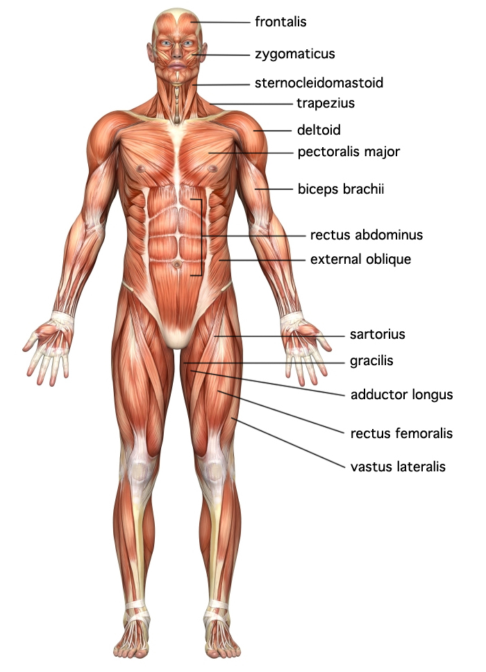
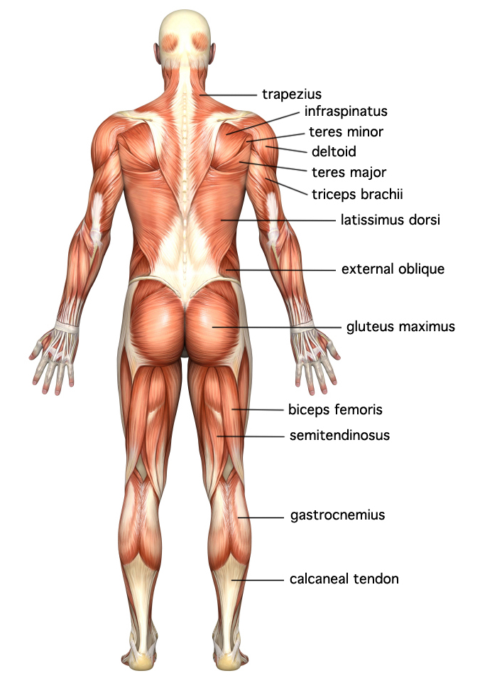
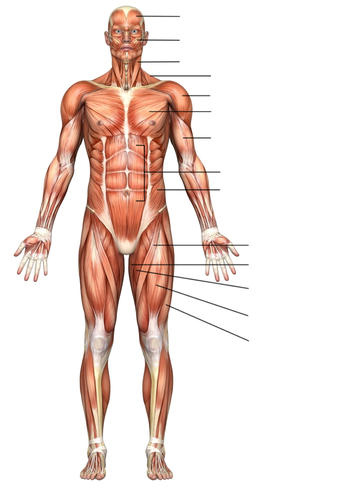
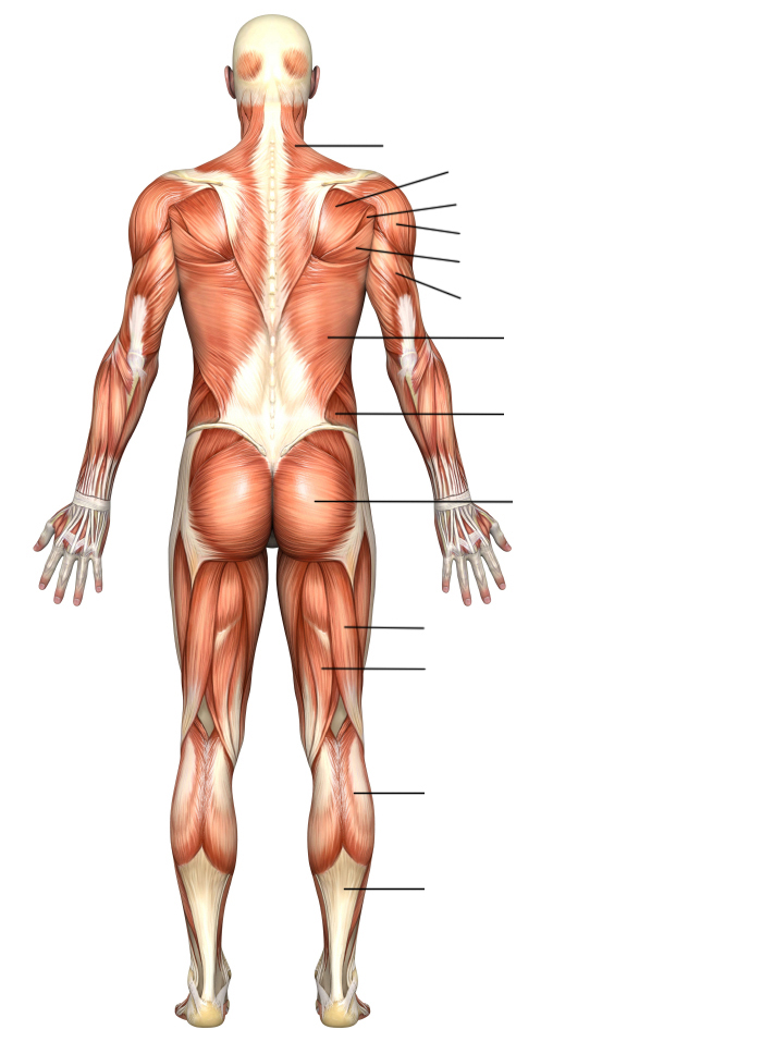
Major Muscles of the Human Body
Review the major muscles of the human body for both anterior and posterior views.
Anterior View
Posterior View
Rigor Mortis
When a person dies, the respiratory and circulatory systems stop delivering oxygen and nutrients to the muscles. Rigor mortis, the stiffening of muscles due to this lack of oxygen and energy, starts to develop about 1-2 hours after death.
Coroners can determine the time of death by knowing how the muscle reactions develop over time.
Initially, stiffening of muscles begins in the upper body and continues through the lower body. Full rigor mortis usually takes between 10 and 12 hours. As time goes on, the stiffening leaves the body in the same order as it progressed—first from the face, neck, and upper body and on to the lower extremities. Rigor mortis disappears completely after approximately 72 hours.
The times of onset and disappearance can vary considerably depending on environmental factors. Coroners must consider these factors when attempting to estimate the times of death.
Checking for Understanding
1. Label the muscles of the body—anterior view.
2. Label the muscles of the body—posterior view.
3. How does the structure of the three types of muscle cells relate to their function?
4. Explain the mechanism for muscle contraction.
5. Why would a disease that causes paralysis of smooth muscles be especially serious?
6. A murder victim is found with relaxed face muscles, but a stiff upper and lower body. How long has it been since the victim died?