


The Circulatory System
Teacher Pages
The human circulatory system is composed of blood, blood vessels and the heart. There are three types of blood vessels which differ in structure and function. The heart serves as the pump for circulating blood around the body. Blood is the fluid that transports nutrients to all body tissues and wastes to the appropriate organ for elimination.
Three types of blood vessels:
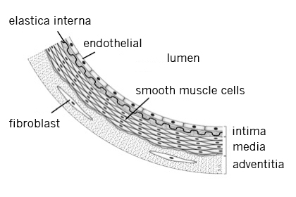
1) Arteries are thick-walled, muscular vessels that almost always carry oxygenated blood away from the heart. The muscular walls allow the arteries to pulse and help push the blood farther along the circulatory pathway as the blood travels away from the heart.
Arteries carry blood from the heart that has picked up a fresh supply of oxygen from the lungs. It is O2-rich, CO2-poor, and bright cherry red.
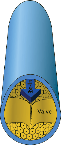 2) Veins are thin-walled and have valves that prevent the blood from flowing back in the wrong direction. Veins carry blood heading toward the heart. Blood coming from the tissues has delivered O2 and picked up CO2 from the cells.
Venous blood is O2-poor, CO2-rich, and dark red.
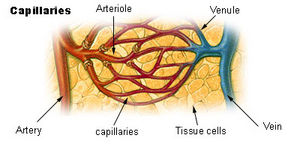 3) Capillaries are blood vessels that are one-celled thick and “connect” arteries with veins. The transfer of nutrients and oxygen from the blood stream into the body tissues takes place in the capillaries where diffusion allows the movement of materials across the capillary membrane and into the tissues.
Heart Structure
The heart is an amazing organ! About the size of a human fist, it pumps blood throughout the body, providing vital nutrients and oxygen to tissues and organs. The process also allows metabolic waste products to be delivered to the appropriate organ for elimination.

Source: http://en.wikipedia.org/wiki/Aorta
The human heart is a hollow organ composed almost totally of muscle. It is surrounded by a protective sac called the pericardium and has three muscular layers. The middle layer, the myocardium, is the largest and does most of the work that makes the heart contract to pump the blood throughout the body.
Structurally, the human heart consists of four chambers and is arranged in right and left halves separated by a thick, muscular wall, the septum. The septum prevents the oxygen-rich blood from mixing with the oxygen-poor blood. (As you learned in your other biological studies, this is not the case in all animals, or even in all vertebrates. Animals whose oxygen-rich and oxygen-poor blood mix have much less efficient circulatory systems.)
Of the four chambers, the upper two chambers or atria, are thin-walled and receive blood into the heart. The lower two chambers, the ventricles, are thick-walled and muscular and are the pumping chambers. Blood leaves the heart from these chambers.
The structure and function of the various blood vessels and the heart is easily understood by following the route of oxygenated and non-oxygenated blood through the body. Since blood circulates through the body, one can start at any point in the route and come back to the same point after completing the route. In this discussion, we will start at the point when blood is returning to the heart from body tissues.
Remember: Arteries carry blood AWAY from the heart. Any time blood is traveling away from the heart, it is in an artery.
Veins carry blood TO the heart. Any time the blood is on its way back to the heart, it is in a vein.
Capillaries are sites where transfer of nutrients and wastes occur.
The Flow of Blood
Blood returns to the heart from the upper body regions via the superior vena cava and from the lower body regions via the inferior vena cava. The transfer of nutrients into the tissues has occurred, so this blood is oxygen-poor and carbon dioxide-rich.
Blood enters the right atrium and flows through the a-v (tricuspid) valve to enter the right ventricle. (Remember, these chambers are thick-walled and muscular to forcefully push the blood out of the heart.)
Blood leaves the heart through the pulmonary valve by way of the right and left pulmonary arteries. (“Pulmonary” refers to the lungs.)
Blood enters the lungs, releases its carbon dioxide, and picks up a fresh supply of oxygen
Blood returns to the heart from the lungs through the right and left pulmonary veins and enters the left atrium
Blood flows through the a-v (bicuspid) valve and enters the left ventricle
Blood leaves the left ventricle through the aortic valve and enters the aorta, the largest artery in the body
Blood travels throughout the body through arteries that branch and become smaller and smaller. Every major organ has its own set of arteries and veins. For example, arteries that “feed” the kidney are called the renal arteries; arteries associated with the liver are called the hepatic arteries.
When the blood enters the capillaries transfer of nutrients and wastes occur. Blood then flows into vessels that thin-walled (veins). Veins gradually become larger as blood begins its route back to the heart.
Eventually, the blood enters the superior and inferior vena cava (the largest veins in the body) again and one circulatory route has been completed.
 Blood
Blood
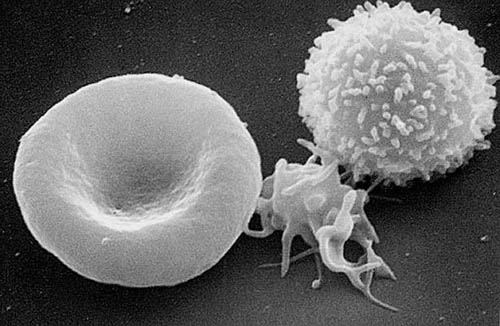
Blood is made up of cells and plasma. The major types of cells are red blood cells, white blood cells, and platelets.
Red Blood Cells
Also called erythrocytes, red blood cells (rbc) are the most numerous cells in the blood. One milliliter of blood normally contains about 5 million rbc’s depending on age and gender. Hemoglobin on the red blood cells combines with oxygen in the lungs. In this way, oxygen is taken around the body and is released to the tissues. Red blood cells are produced in the bone marrow and last approximately 20- 120 days. Thus, bone marrow must constantly produce new red blood cells.
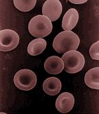 Mature red blood cells look like flattened disks that are thinner in the middle than around the edges. A variety of diseases and disorders can be diagnosed by looking at the appearance of red blood cells. Toxic chemicals and snake
  venom can distort the appearance of red blood cells and parasites such as those that cause malaria and blackwater fever can destroy the cells. Hemolytic anemia (when the cells burst and there are not enough rbc’s in the body) results from each of these.
White Blood Cells
Leucocytes, or white blood cells (wbc) are present in far fewer numbers than red blood cells. White blood cell counts are normally 5,000 –10,000 mm3. Elevated counts can indicate infection because these cells guard against infection, fight parasites and attack bacteria. Some wbc’s do this by phagocytosis, or engulfing the bacteria or other foreign cells in the blood. Other white blood cells leave the blood and enter the tissues to engulf invading organisms and still others release chemicals that help the body fight disease.
The different types of white blood cells not only have different functions but they can indicate different disorders or conditions. Blood counts and the appearance of various cells are used as diagnostic tools to identify many different conditions.
Note: See Table— Comparison of Types of White Blood Cells for microscopic images, diagrams, normal amounts, and size information.
 Platelets
Platelets and plasma proteins work together to clot blood when an injury has occurred. Platelets in the blood come into contact with torn edges of the blood vessel. Their surfaces become sticky and clotting proteins in the plasma start a series of complex chemical reactions. A sticky mesh forms over the wound or injury and the clot forms.
Clotting is extremely important to health and welfare. Platelet counts and plasma protein levels are diagnostic tools for a number of clotting disorders and are also measured when patients must undergo therapeutic anti-coagulant treatment.
Plasma
You have just read about the solid part of the blood. Cells make up about 45% of the volume of the 4-6 liters of blood in the human body. The other 55% of the volume is the liquid part of the blood, the plasma. Plasma is a light, sticky straw-colored liquid that is 90% water. Salts, sugars, dissolved fats, proteins, nutrients, hormones, clotting factors, and waste products are carried in the plasma.
Blood Pressure
Blood pressure is produced by the force of the heart pumping blood away from the heart and through arterial walls. Muscular arterial walls also produce pressure. Therefore, when we measure blood pressure we are measuring the pressure of the blood against the inside of the artery walls. When the heart relaxes within the beat, the pressure against the artery walls decreases, but it is still present.
Pressure is measured by a sphygomanometer. This instrument measures blood pressure by pumping air into a cuff positioned around an artery. The cuff presses against the artery and measures the pressure exerted against it by the blood pumped by the heart and arteries.
Blood pressures consist of two measurements. One is the systolic pressure—the pressure in the arteries when the ventricles contract. The other is the diastolic pressure—the pressure of the blood felt in the arteries when the ventricles are relaxed. Normal adult blood pressure is 120/80 mmHg..
Hypertension, or high blood pressure, can damage heart muscle over time. Heart disease occurs and the risk of heart attack or stroke increases.
Pulse
Arteries, unlike veins, are muscular blood vessels. They help push the blood along the circulatory route to keep blood flowing through the body at more constant rate. Arteries pump as the heart pumps and the pulse that results can be felt in arteries near the surface of the skin.
Pulse rates are measured in beats per minute. Normal adult pulse rates average 60-80 beats per minute.
Checking for Understanding
- Describe the flow of blood through the circulatory system.
See the description above.
- What are the major differences among the three types of blood vessels? Complete the following table.
|
Type of Blood Vessel |
Function |
Structure of Walls |
Distinguishing Feature |
|
Arteries |
Carry blood AWAY from the heart
|
Thick-walled, muscular |
Pulse with heartbeats |
|
Veins |
Carry blood TOWARD the heart
|
Thin-walled |
Have valves to prevent backflow of blood |
|
Capillaries |
Connect arteries and veins in the flow of blood in the body
|
Single-celled walls |
Site of transfer of nutrients and wastes to and from the tissues |
- Why can blood cells be used to diagnose certain disease conditions?
Blood cells have different structures and functions within the body. When certain disease conditions arise, they can cause a change in number or appearance of the cell involved in a function related to the disease.
- What does an elevated white cell count indicate and why?
White blood cells fight infection. Therefore, more white blood cells are produced by bone marrow in response to disease conditions. A normal wbc count is 5,000 to 10,000/mm3. A white blood cell count over 10,000 mm3 indicates an infection that may not be outwardly visible.
- Name several conditions that will cause a change in red blood cells.
Toxic chemicals, snake venom, and parasites such as those that cause malaria and blackwater fever.
- Describe what is happening in the diastolic and systolic phases of a heartbeat.
Blood pressures consist of two measurements. One is the systolic pressure—the pressure in the arteries when the ventricles contract. The other is the diastolic pressure—the pressure of the blood felt in the arteries when the ventricles are relaxed. Normal adult blood pressure is 120/80 mm Hg..
- Compare and contrast the structure and function and normal numbers of the three major types of blood cells. Complete the following table.
|
Type of Blood Cell |
Function |
Normal Count |
|
White blood cell |
Fights infection |
5,000-10,000 mm3 |
|
Red blood cell |
Carries oxygen to the tissues |
5 million/mm3 |
|
Platelet |
Helps clot blood at injury sites |
150,000-400,000 mm3 |
- Identify the following as having either O2-rich or CO2-rich blood:
a) aorta—O2 rich
b) coronary arteries—O2-rich
c) inferior vena cava—CO2-rich
d) coronary veins—CO2-rich
e) left atrium—O2-rich
f) right atrium—CO2-rich
g) left ventricle— O2-rich
h) right ventricle— CO2-rich
i) superior vena cava—CO2-rich
- Almost all arteries carry oxygenated blood. What is the one exception?
Pulmonary arteries carry blood away from the right ventricle. This blood has just returned from body tissues and is oxygen-poor and carbon dioxide-rich. The pulmonary arteries deliver the blood to the lungs.
10, Identify the cells in the blood smear:
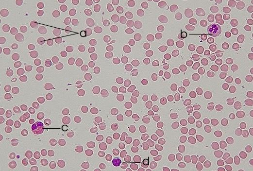
http://en.wikipedia.org/wiki/Image:Blood_smear.jpg
a) red blood cell
b) neutrophil
c) eosinophil
d) lymphocyte
e) platelet
11. Think back to your studies of cell structures. Why do you think cells of heart muscle contain so many mitochondria?
Mitochondria are cell structures that produce ATP through the process of cellular respiration. Heart muscles cells are very active all the time and require constant supplies of energy.


